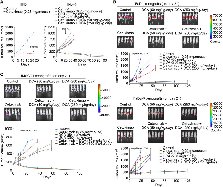
Figure 7. Cetuximab plus PDK1 inhibition induces apoptosis in HNSCC cells with acquired resistance to cetuximab and inhibits their growth in vivo when used in combination with DCA.
(A) Left, established HN5 xenografts (~250 mm3) were untreated or treated with cetuximab (0.25 mg/mouse, twice a week for 3 weeks). Right, HN5-R xenografts (~250 mm3) were untreated or treated with the same dose of cetuximab, DCA (50 or 250 mg/kg/day), or DCA plus cetuximab for 3 weeks. (B) Established FaDu (upper) and FaDu-R (lower) xenografts (~250 mm3) were untreated or treated as described in A for HN5-R xenografts. After completion of treatments, the mice were subjected to IVIS imaging and then observed for additional days for tumor progression or regression. The recording of tumor sizes was stopped when fewer than 4 mice remained in any group because of sacrifice of mice with large tumor burden or morbid or moribund status. (C) Established UMSCC1 xenografts (~150 mm3) were untreated or treated with cetuximab (0.25 mg/mouse, twice a week for 3 weeks), DCA (50 or 250 mg/kg/day), or DCA plus cetuximab for 3 weeks. After completion of treatments on day 21, the mice were subjected to IVIS imaging and then observed for additional days for tumor progression or regression. The recording of tumor sizes was stopped when fewer than 4 mice remained in any group because of sacrifice of mice with large tumor burden or morbid or moribund status. Error bars indicate ± SEM. Rx, treatment.

