
Fig 2. 3D structure of scFv-108.
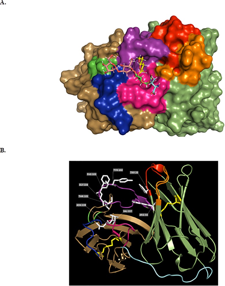
Panel a shows the potential site of interaction between scFv-10D8 and its ligand, β-D-galactofuranose(1–4)N-acetylglucosamine. Model obtained in silico with the most stable interaction between the two molecules using Hex 8.0 software. β-D-galactofuranose(1–4)N-acetylglucosamine is represented by the sticks model, while scFv-10D8 is represented by the surface model for better visualisation of the antigen-binding site. Panel b represents scFv-10D8 in a VH-linker-VL format, highlighting the β-sheets, turns and flexible regions. In yellow are the cysteine residues and the disulfide bonds that are critical for the antibody structure.