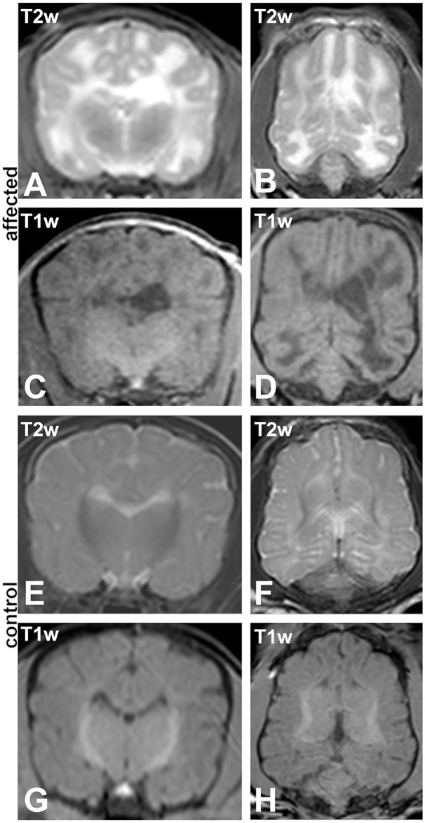
Fig 1. Magnetic resonance imaging (MRI).
(A-D) Affected 4 weeks old Schnauzer puppy (no. 1) and (E-H) a clinically normal sibling at the same age (no. 14). (A, B, E, F) T2 weighted (T2w) sequences; (A, E) transversal and (B, F) dorsal. (C, D, G, H) T1 weighted (T1w) sequences; (C, G) transversal and (D, H) dorsal. MRI reveals mildly enlarged and asymmetric lateral ventricles and a T2w-hyperintense/T1w-hypointense lesion diffusely involving the whole cerebral white matter in affected animals. The age matched unaffected sibling shows beginning myelination in the central deep white matter, typical for the age.

