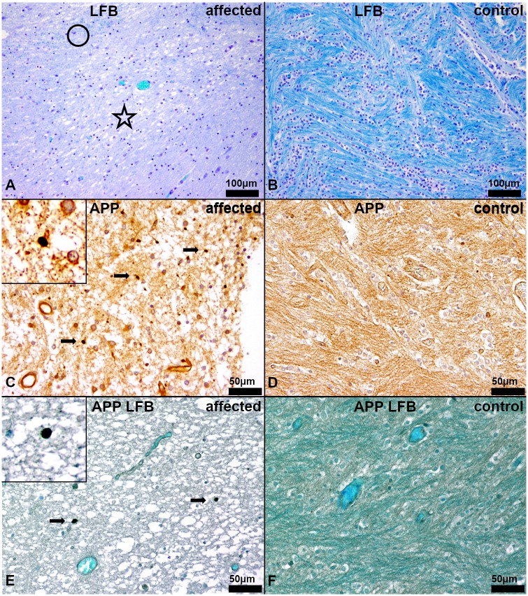
Fig 4. Histochemistry and immunohistochemistry of the white matter in the centrum semiovale of the cerebrum.
(A, C, E) Affected puppy no. 1 and (B, D, F) age matched healthy control no. 1009. (A) Severe, diffuse lacking bluish staining, indicating myelin loss and edema in the centrum semiovale (asterisk). Only single fine strands of myelin can be detected (circle). (LFB). (B) Regularly developed myelin characterized by prominent bluish staining in the white matter (dark blue). (LFB). (C) Areas of severe myelin loss in the diseased puppies reveal low numbers of damaged axons (arrows). Vacuolization and loosening of the parenchyma indicate moderate edema. Inset shows a damaged axon at a higher magnification. (β-amyloid precursor protein, APP). (D) No axonal damage is detectable in healthy control animals. (APP). (E) Severe, diffuse lacking blue-greenish staining, indicating myelin loss and edema, in the centrum semiovale, containing low numbers of damaged axons (arrows). Vacuolization and loosening of the parenchyma represent the moderate edema. Inset shows a damaged axon at a higher magnification. (APP, LFB). (F) Regularly developed myelin characterized by prominent blue-greenish staining in the white matter (dark teal) without any damaged axons. (APP, LFB).

