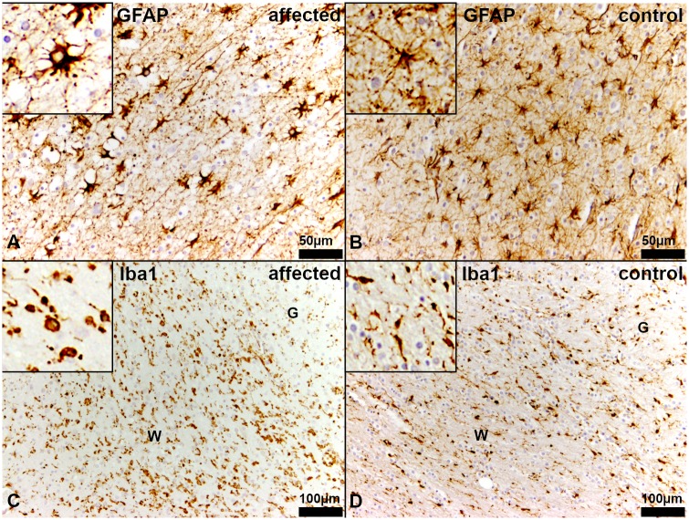
Fig 7. Immunohistochemistry of the white matter in the centrum semiovale of the cerebrum.
(A, C) Affected puppy no. 1 and (B, D) age matched healthy control no. 1009. (A) There is a reduced density of astrocytes in the white matter of affected puppies. Most of the astrocytes have a plump morphology developing few short and bulky processes. Inset shows an astrocyte at a higher magnification. (GFAP). (B) In the white matter of age matched control puppies, astrocytes have long, slender branching processes. Inset shows an astrocyte at a higher magnification. (GFAP). (C) Diseased Schnauzer puppies reveal an elevated number of macrophages/microglia in the white matter. Many of these macrophages/microglia have an amoeboid or reactive morphology (arrows). Inset shows amoeboid macrophages/microglia at higher magnification. (Iba1). (D) In healthy control dogs, there is an equal distribution of microglia in the white (W) and the grey (G) matter. Most of the macrophages/microglia represent the ramified, non-reactive type. Inset shows macrophages/microglia at a higher magnification. (Iba1).

