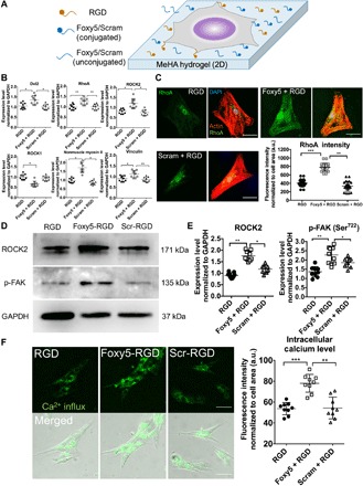
Fig. 2. Foxy5 peptide–conjugated hydrogel scaffolds activate noncanonical Wnt signaling to trigger RhoA signaling and elevate intracellular calcium.

(A) Schematic illustration of the seeding of hMSCs on the Foxy5/Scram + RGD peptide–functionalized 2D hydrogel substrate. (B) Gene expression level of the RhoA signaling cascade (Wnt5a coreceptor Dvl2, RhoA, ROCK), downstream mechano-effector (NMII), and major focal adhesion adaptor protein (vinculin) in hMSCs in 3D porous hydrogels conjugated with RGD peptide alone (RGD), Foxy5 and RGD peptide (Foxy5 + RGD), or scrambled Foxy5 peptide and RGD peptide (Scram + RGD), respectively, after 7 days of osteogenic culture (n = 9). (C) Representative micrographs of fluorescence staining for F-actin (red), nuclei (blue), and RhoA (green) in hMSCs cultured on the 2D RGD, Foxy5 + RGD, and Scram + RGD hydrogels. Quantification showed a significantly higher RhoA staining intensity in the Foxy5 + RGD group than in the RGD and Scram + RGD groups (n = 20). a.u., arbitrary units. (D) Western blot bands and quantification of the expression level of mechano-responsive kinases ROCK2 and p-FAK (phosphorylated at the Ser722 sites) in each group (RGD, Foxy5 + RGD, Scram + RGD). (E) Representative merged fluorescence and bright-field micrographs of intracellular calcium in hMSCs cultured on RGD, Foxy5 + RGD, and Scram + RGD 2D hydrogels (stained with Fura-AM). (F) Quantification showed a significantly higher intracellular calcium level in the Foxy5 + RGD group than in the RGD and Scram + RGD groups. Scale bars, 50 μm. Data are shown as the means ± SD (n = 9). Statistical significance: *P < 0.05, **P < 0.01, and ***P < 0.001.
