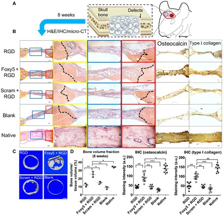
Fig. 5. Functionalization of biomaterial scaffolds with the Wnt5a mimetic peptide substantially enhances the in situ regeneration of integrated and mature bone tissues.

(A) Schematic illustration of the implantation of rMSC-seeded and peptide-functionalized porous hydrogels in rat calvarial defects. (B) H&E staining and immunohistochemical staining of the native healthy bone tissue and the calvarial defects treated with the RGD hydrogels, Foxy5 + RGD hydrogels, Scram + RGD hydrogels, and no hydrogels (blank) 8 weeks after implantation (n = 3). High-magnification images showing the defect/native bone boundaries highlighted in yellow and red boxes and defect center areas in blue boxes in the low-magnification images of H&E-stained sections. The dotted lines indicate the boundary between the defect and native bone. The newly formed bone was seamlessly integrated with the neighboring native bone in the Foxy5 + RGD group. Scale bars, 50 μm. (C) Top view of 3D micro-CT images showing calvarial bone defects after 8 weeks in all groups (n = 3). (D) Bone volume (normalized to total tissue volume, BV/TV) in the calvarial defects in all groups after 8 weeks (n = 3). The bone volume of healthy rat calvarial bone is shown as the benchmark. Quantification of the immunohistochemical staining intensity of the osteogenic markers, including osteocalcin and type I collagen, showing the higher intensity in the Foxy5 + RGD group compared with those of the RGD and Scram + RGD control groups. Data are shown as the means ± SD (n = 9). Statistical significance: *P < 0.05, **P < 0.01, and ***P < 0.001 significant difference.
