
Fig. 5. PP2Cδ negatively regulates basal and UV damage–induced ATM phosphorylation.
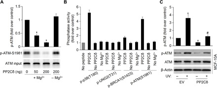
(A) PP2Cδ dephosphorylates intact ATM at phospho-Ser1981. An in vitro phosphatase assay was performed by incubating immunoprecipitated ATM and purified PP2Cδ, followed by Western blot probing with antibodies to ATM phospho-Ser1981. The data represent mean ± SD from three separate experiments. *P < 0.05 versus control. (B) Phosphopeptides from p38 MAPK (positive control), UNG2 (negative control), BRCA1 (phospho-Ser1423), and ATM (phospho-Ser1981) were incubated with PP2Cδ in an in vitro phosphatase assay. Release of free phosphate was measured by absorbance at 630 nm in the presence of molybdate dye. Reactions were also carried out without magnesium or peptide. (C) MCF-10A cells were transfected with EV or plasmid expressing WT PP2Cδ, followed by UV treatment (20 J/m2 for an 8-hour recovery). Cells were lysed, and the ATM phosphorylation and protein levels were detected by Western blotting with specific antibodies. The data represent mean ± SD from three separate experiments. *P < 0.05 versus EV/UV (−); #P < 0.05 versus EV/UV (+).