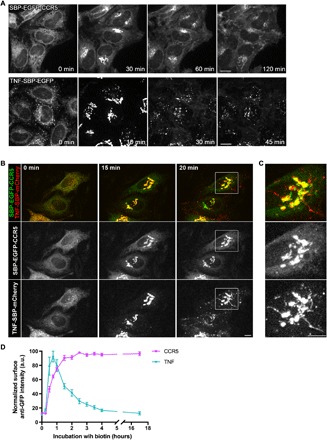
Fig. 1. Differential anterograde transport of CCR5 and TNF.

(A) Synchronized transport of CCR5 (top) and TNF (bottom) in HeLa cells stably expressing Str-KDEL_SBP-EGFP-CCR5 or Str-KDEL_TNF-SBP-EGFP. Trafficking was induced by addition of biotin at 0 min. Scale bar, 10 μm. (B) Dual-color imaging of the synchronized transport of SBP-EGFP-CCR5 and TNF-SBP-mCherry transiently coexpressed in HeLa cells. Streptavidin-KDEL was used as an ER hook. Release from the ER was induced by addition of biotin at 0 min. Scale bar, 10 μm. (C) Magnification (×2.8) of the Golgi complex region is displayed. Scale bar, 10 μm. (D) Kinetics of arrival of CCR5 (magenta) or TNF (cyan) to the cell surface after release from the ER measured by flow cytometry. Ratio of cell surface signal divided by GFP intensity was used for normalization. a.u., arbitrary units. The mean ± SEM of three experiments is shown. See also movies S1 to S3.
