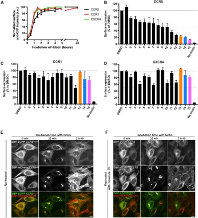
Fig. 3. Three small molecules inhibit the transport of CCR5 and do not affect transport of CCR1 or CXCR4.

(A) Kinetics of synchronized transport of three chemokine receptors—CCR5 (black), CCR1 (red), and CXCR4 (green)—using the RUSH assay. Trafficking was induced by addition of biotin at time 0. Surface expression of CCR5, CCR1, and CXCR4 was measured by flow cytometry using an anti-GFP antibody. Ratio of cell surface signal to GFP intensity was used for normalization. The mean ± SEM of three experiments is shown. End-point measurement (2 hours) of the effects of the hit molecules on the trafficking of CCR5 (B), CCR1 (C), and CXCR4 (D) in HeLa cells. Cells were pretreated for 1.5 hours with molecules at 10 μM. Cargo present at the cell surface 2 hours after release from the ER was measured by flow cytometry using an anti-GFP antibody. Ratio of cell surface signal to GFP intensity was used for normalization. The mean ± SEM of three experiments is shown. Real-time synchronized secretion of CCR5 and CCR1 was monitored using dual-color imaging in nontreated HeLa cells (E) and in HeLa cells pretreated for 1.5 hours with 10 μM molecule 13 (F). Scale bars, 10 μm.
