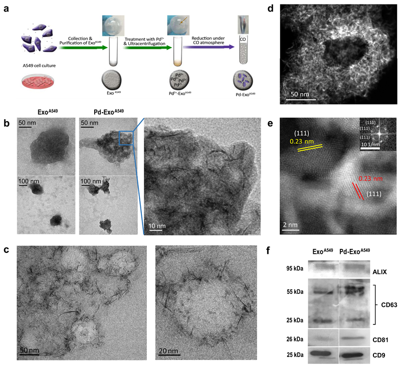
Figure 1. Preparation and characterization of Pd-functionalised exosomes.
(a) Overview of the step-wise protocol to generate Pd-ExoA549 from A549 cells. (b) Representative TEM images of ExoA549 and Pd-ExoA549 at different magnifications. (c) Representative cryo-TEM images of Pd-ExoA549 at two magnifications. (d) HRSTEM-HAADF image of a representative Pd-ExoA459. (e) HRSTEM-HAADF zoomed-in image showing highly-crystalline Pd nanostructures with a lattice spacing of 0.23 nm, which correspond to Pd (1 1 1)-surfaces. Inset shows the FFT spectrum generated from the image. (f) Western blots of exosome-specific biomarkers ALIX, CD63, CD81 and CD9 of ExoA549 and Pd-ExoA549.

