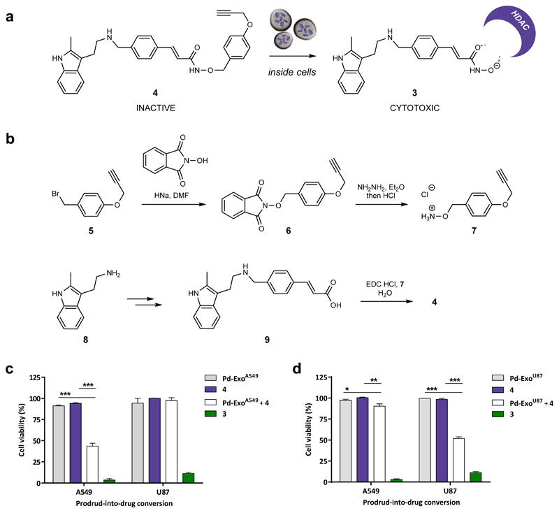
Figure 4. Design and synthesis of prodrug 4 and targeted intracellular activation mediated by Pd-ExoA549 and Pd-ExoU87 in A549 and U87 cells.
(a) Intracellular Pd-ExoA549-mediated conversion of prodrug 4 into clinically-approved HDAC inhibitor 3. (b) Total synthesis of compound 4. (c) Pd-ExoA549-mediated uncaging of 4 inside cells. A549 and U87 cells were incubated for 6 h with 0.4 and 0.53 μg / 100 μL, respectively. Cells were thoroughly washed to eliminate extracellular vesicles followed by addition of prodrug 4 (0.2 μM). Controls: Pd-ExoA549 only (–ve control, grey); prodrug 4 only (–ve control, purple); 3 (+ve control, green). Cell viability was measured at day 5 using PrestoBlue. Error bars: ± SEM, n = 3. (d) Pd-ExoU87-mediated uncaging of 4 inside cells. A549 and U87 cells were incubated for 6 h with 0.4 and 0.53 μg / 100 μL, respectively. Cells were thoroughly washed to eliminate extracellular vesicles followed by addition of prodrug 4 (0.2 μM). Controls: Pd-ExoU87 only (–ve control, grey); prodrug 4 only (–ve control, purple); 3 (+ve control, green). Cell viability was measured at day 5 using PrestoBlue. Error bars: ± SEM, n = 3.

