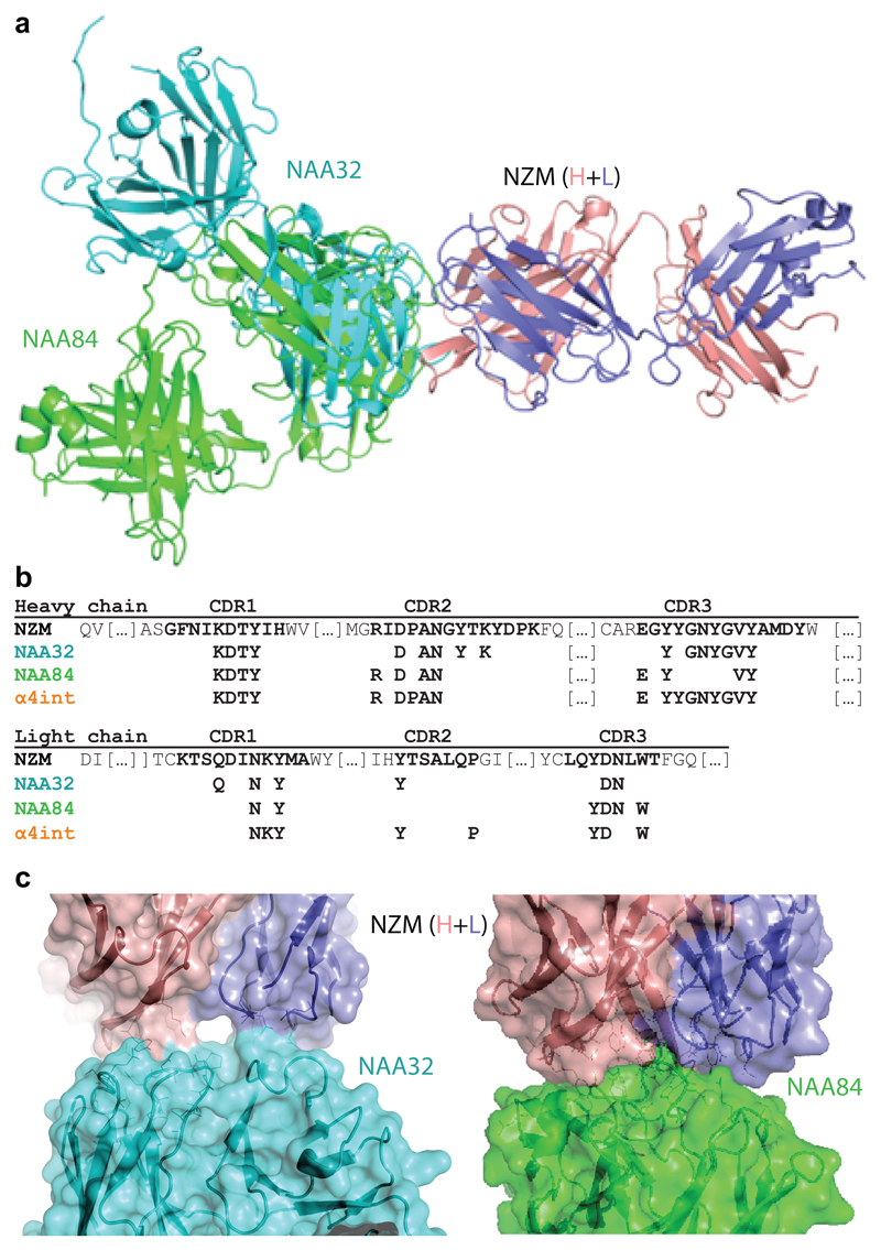
Fig. 3. Structural features of the interaction of NZM with a NAb and a BAb.
a, Superimposition of the antigen-binding fragment of NZM in complex with NAA84 (NAb, green) and NAA32 (BAb, cyan). NZM heavy and light chains are shown in salmon and slate blue, respectively. Proteins are displayed in ribbon diagram. b, Alignment of the NZM residues that are recognized by NAA32, NAA84 and α4 integrin. c, Detailed visualization of the interacting interfaces of NZM and NAA32 (left) and NAA84 (right). The antibodies are shown as ribbon diagrams with overlapping surfaces. The Sc values were 0.696 and 0.707 for NAA32/NZM and NAA84/NZM complexes, respectively.

