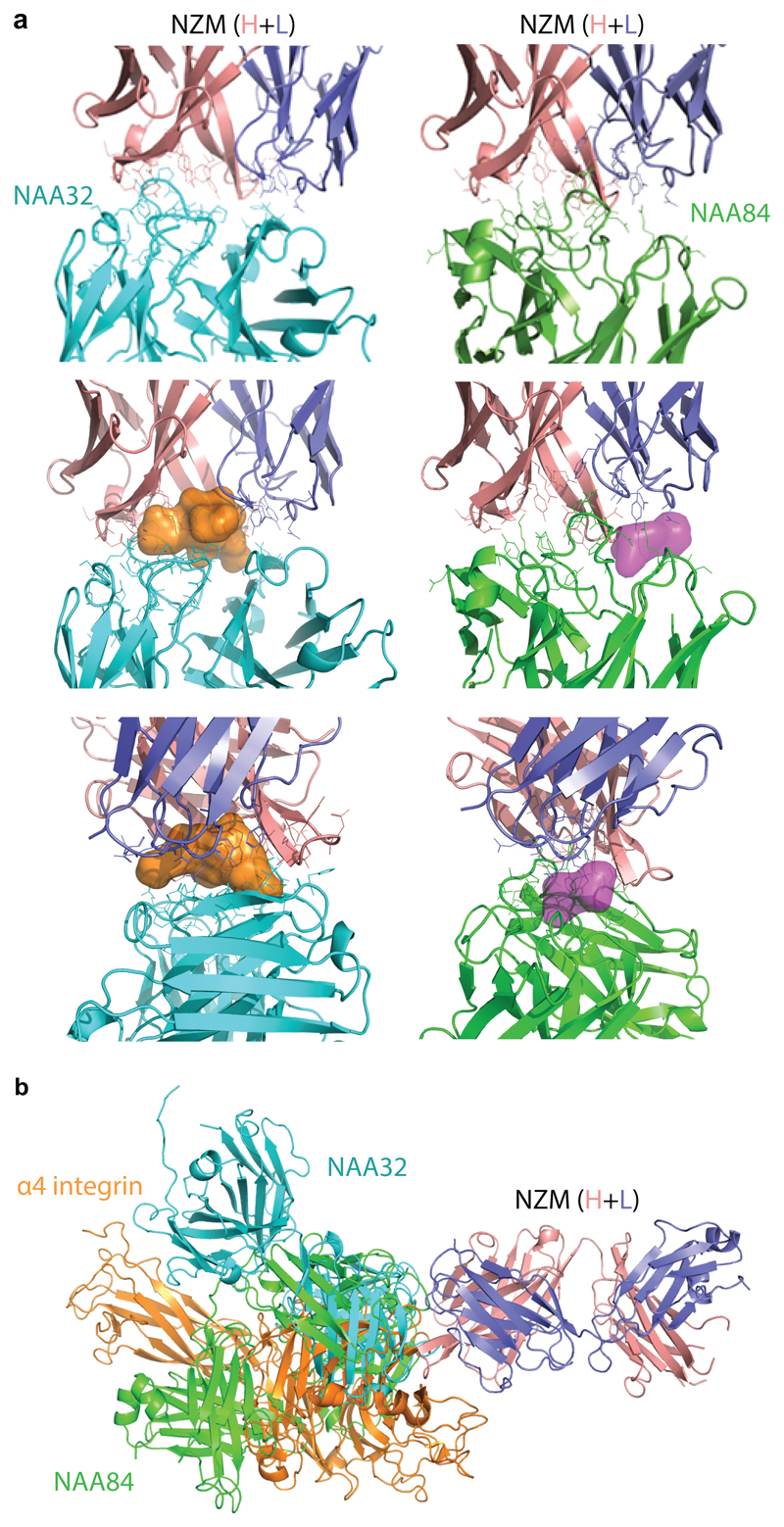
Extended Data Fig. 2. Structural details of the interaction of NZM with a NAb, a BAb and α4-integrin.
a, Closer view of the interaction interface between NZM and NAA32 (left) and NAA84 (right). Epitope and paratope residues are shown in solid sticks. Proteins are displayed in ribbon diagram. The empty space in the interface between the NZM and NAA32 or NAA84 is represented as orange or purple surface, respectively, in two different orientations b, Superimposition of the antigen-binding fragment of NZM in complex with NAA84 (NAb, green), NAA32 (BAb, cyan) and α4-integrin (orange). NZM heavy and light chains are shown in salmon and slate blue, respectively. Proteins are displayed in ribbon diagram.

