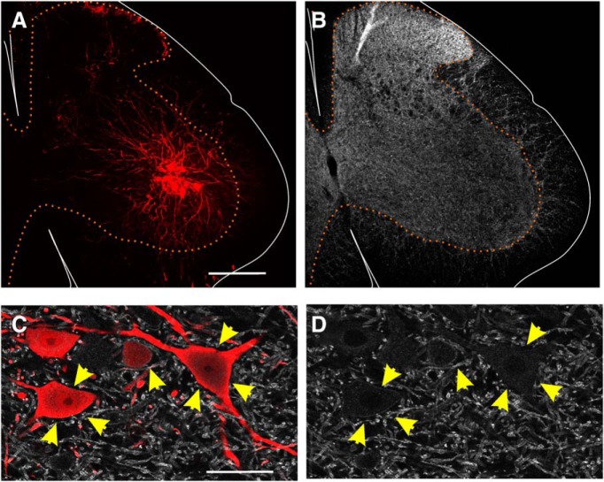
Figure 4.
KCC2 protein is not lost on motoneuron somata or dendrites 3 d after sciatic nerve cut/ligation. A, B, AAV1-mCherry filled LG motoneurons (A) and KCC2-IR (B) in the same section 3 d following sciatic nerve cut/ligation. Scale bar = 200 μm C, D, Representative high-magnification (60× 1) images of the motoneurons (red) above and the KCC2-IR (white) on their somata and dendrites (yellow arrows). There is minimal loss of KCC2-IR on both the soma and dendritic processes. No loss is visible on mid to distal dendrites. Scale bar = 50 μm.

