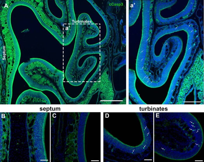
Figure 3.
Cell death in the OE in P25 mice. A, Semipanoramic view of the OE including the septum and turbinates. a′, Caspase 3+ cells (green) are mainly spread across the turbinates (arrows). Almost no caspase 3+ cells were observed along the septal wall. B, C, High magnification of the septal wall showing the absence of caspase 3+ cells. D, E, High magnification of different turbinates showing the presence of several caspase 3+ cells (arrows). Scale bars: A, 400 μm; a′, 200 μm; B–E, 50 μm.

