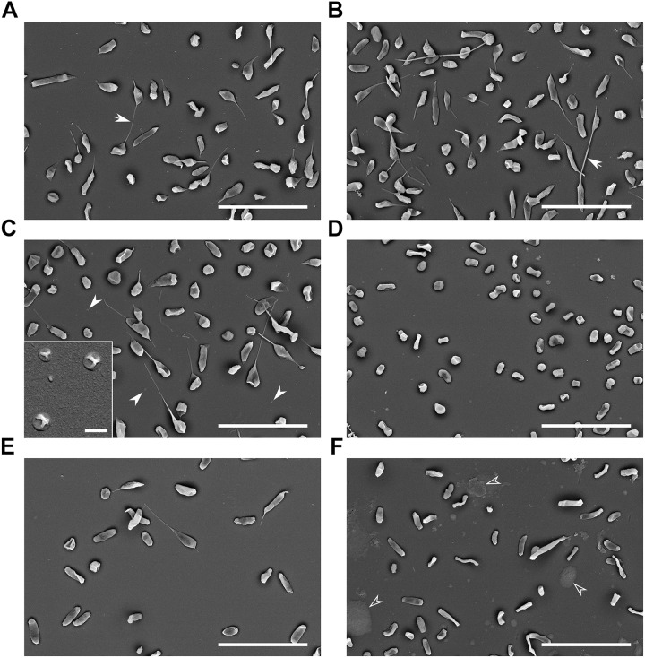
FIGURE 2.
Scanning electron microscopy of bacteria cultured under various conditions. (A) Control: BHI, 37°C, pH 6.8, 16 h, arrows point to the tubular protrusions connecting adjacent cells; (B) oxidative stress: 10 mM H2O2; (C) 42°C, full arrowheads point to the small spherical vesicles on the background, inset: higher magnification of these small spherical vesicles; (D) 25°C; (E) pH 5.3; and (F) agar plate grown bacteria, empty arrowheads point to the unspecified material. Bars in main images: 5 μm; C-inset: 200 nm.

