FIGURE 2.
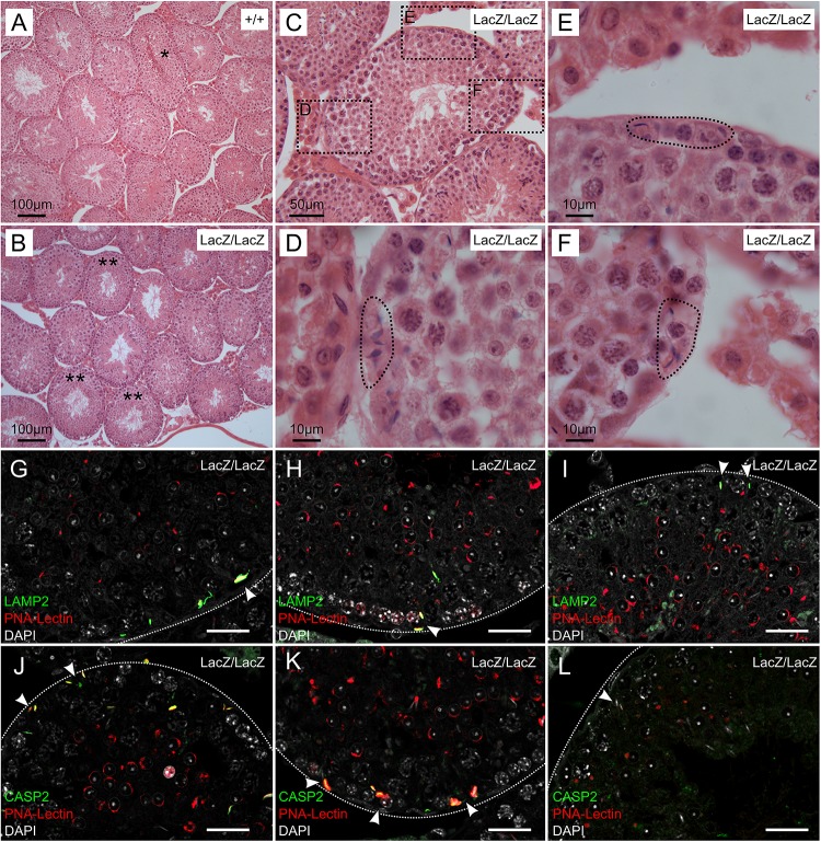
Immunochemical staining of caspase 2 and LAMP2 in Fbxo7LacZ/LacZ testis sections. (A–B) Low magnification view of H&E sections from wild type (A) and Fbxo7LacZ/LacZ testes (B). ∗ In panel (A) indicates a tubule at stage VII-VIII with sperm heads lined up at the lumen awaiting release. These were never observed in Fbxo7LacZ/LacZ testes. ∗∗ In panel (B) indicates tubules with a layer of round spermatids but which lack elongating spermatids. (C) High magnification view of a Fbxo7LacZ/LacZ tubule lacking elongating spermatids: (D–F) Close up zoom from panel C at the indicated locations. The dotted outlines indicate “graveyards” containing multiple phagocytosed spermatid heads. (G–I) Immunofluorescent stains in Fbxo7LacZ/LacZ tubules for LAMP2 (green) with PNA-lectin (red) to stage the tubules and DAPI counterstain (gray). Phagocytosed cells marked by LAMP2 were visible at tubule stage VI as indicated by the extent of the lectin-stained acrosomal cap. (J–L) Immunofluorescent stains in Fbxo7LacZ/LacZ tubules for CASP2 (green) with PNA-lectin (red) to stage the tubules and DAPI counterstain (gray). Apoptotic cells marked by CASP2 were visible at tubule stage VI as indicated by the extent of the lectin-stained acrosomal cap. At tubule stage IV (L), occasional mis-localised elongating spermatids were seen next to the basement membrane. These cells were not marked with CASP2 at this stage. For quantitation of spermatid mis-localisation, see Figure 3.

