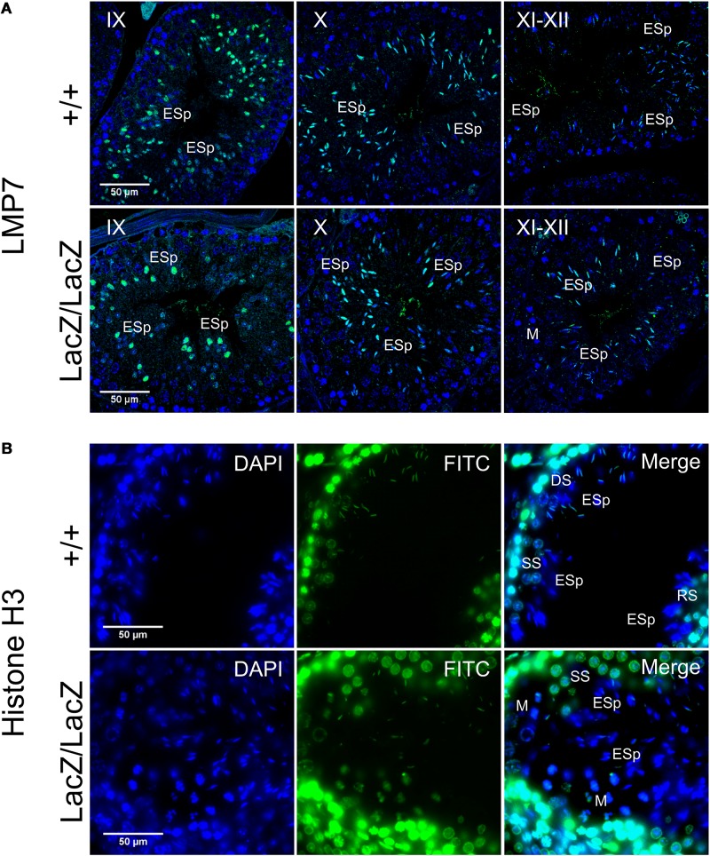
FIGURE 5.
Immunochemical staining of LMP7 and histone H3 in Fbxo7LacZ/LacZ testis sections. (A) Immunochemical staining of LMP7 (FITC, green) in wild type and Fbxo7LacZ/LacZ testes with DAPI (blue) nuclear counterstain. Roman numerals indicate tubule stage. In both genotypes, nuclear LMP7 expression is first seen in early ES at mid-stage IX as the nuclei begin to elongate. This nuclear expression is highest at stage X, and then is lost during stage XI-XII as nuclei complete elongation. (B) Immunochemical staining of histone H3 (FITC, green) in wild type and Fbxo7LacZ/LacZ testes with DAPI (blue) nuclear counterstain. The wild type tubule shown is in transition between stages, with early stage XII (DS next to ESp) at upper left, mid stage XII (SS next to ESp) at lower left and stage XII/I border (M and RS next to ESp) at lower right. Nuclear H3 signal in ESp is still present at early stage XII, is restricted to the posterior of the nucleus in mid stage XII, and entirely lost by stage I. The Fbxo7LacZ/LacZ tubule shown is in mid stage XII, and the H3 signal in the ESp is absent or restricted to the posterior extremity of the nucleus, confirming the kinetics and spatial pattern of H3 removal are indistinguishable between genotypes. Key: DS = diplotene spermatocytes, SS = secondary spermatocytes, M = metaphase figures, RSp = round spermatids, ESp = elongating spermatids.

