Figure 2.
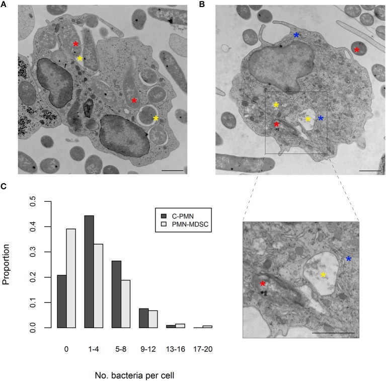
PMN-MDSCs phagocytose bacteria. (A,B) EM images of (A) C-PMN and (B) PMN-MDSC isolated from tumor-bearing dogs. Blue asterisks indicate dilated endoplasmic reticulum, yellow asterisks indicate phagolysosomes, and red asterisks indicate E. coli. Scale bar = 1 μm. (C) Bar graph depicting the proportion of total cells of each cell type analyzed by EM that had the respective range of bacteria internalized. C, cancer; PMN, polymorphonuclear cell; EM, electron microscopy.

