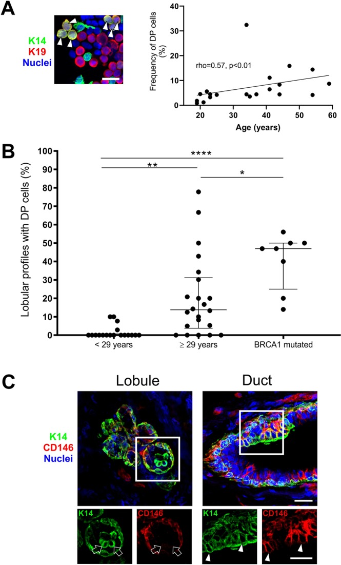
Figure 5.

Variant DP cells accumulate in lobules with age and in tissue from BRCA1 mutation carriers. (A) Immunofluorescent staining of crude smears with K14 (green), K19 (red) and nuclei (blue) (left image). Arrowheads mark DP cells. Bar, 20 µm. A positive correlation was found between age and the frequency of DP cells (right), analyzed by Spearman rank test (rho = 0.57, p < 0.01). (B) Dot plots of the proportion of lobular structures with DP cells in women <29 years (average age: 19.7 years, median: 19), ≥29 years (average age: 44.4 years, median: 43) and BRCA1 mutation carriers (information about donor age not available). The age-selected data for lobules are derived from Supplementary Table S1, which are also included in Fig. 1A. The proportion of lobules containing DP cells is significantly higher in the older age group (20.5%) and in women with BRCA1 mutations (40.5%) as compared to the younger age group (2.9%). *p < 0.05, **p < 0.005, and ****p < 0.000005 using ANOVA with Tukey’s HSD test. Each scatter dot plot is lined at median with interquartile range (<29 years: n = 18, ≥29 years: n = 22, BRCA1 mutated: n = 8). (C) Immunofluorescent staining demonstrating a segregation of DP and CD146+ cells in a lobule (left image) compared to a duct (right image) with CD146-expressing DP cells. K14 is shown in green, CD146 in red and nuclei in blue. Image subsets are shown in single color channels. Arrows indicate DP cells that are CD146neg while arrowheads point to CD146+ DP cells. Bars, 25 µm.
