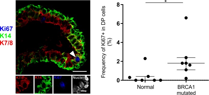
Figure 6.
DP cells are more frequently cycling in tissue from BRCA1 mutation carriers. (A) Normal tissue from a woman with known BRCA1 mutation immunostained for K14 (green), cell cycle marker Ki67 (blue) and luminal keratin marker CAM5.2 (red). Arrowhead marks a Ki67+ DP cell. Bar, 25 µm. Lower panel image subsets are shown in single color channels, including DAPI nuclear stain (white). Bar: 10 µm. (B) Dot plot comparing the proportion of Ki67+ cells in the DP population between women with BRCA1 mutations and women with no known mutations. *p < 0.017 (normal: n = 7; BRCA1 mutated: n = 7) by Mann Whitney test.

