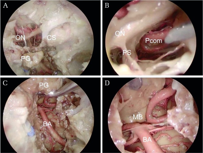
Fig. 1.

Dissection around the pituitary gland via endoscopic transnasal skull base approach in the NVP-embalmed cadavers (No. 3 in Table 1). (A) The pituitary gland (PG), the cavernous sinus (CS), and the optic nerves (ON) are visible after standard transsphenoidal approach and transtuberculum sellae approach. (B) Deep in the suprasellar area, the posterior communicating artery (Pcom) and the pituitary stalk (PS) are under direct vision. (C) Transposition of the pituitary gland and clivectomy provides exposure of full length of the basilar artery (BA). (D) Deeply, the bilateral mammillary bodies (MB) can be observed. Injection of blue silicone was insufficient probably because of insufficient irrigation and removal of thrombi inside the vessels before silicone injection, or hypoplasty of the internal jugular vein. NVP: N-vinyl-2-pyrrolidone.
