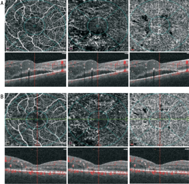
Figure 2. OCTA images of the SCP (A and B, left panel), DCP (A and B, middle panel) and CC (A and B, right panel).
At baseline (A) vessel rarefaction surrounding the foveal avascular zone in the SCP and DCP, microaneurysms and diffuse vessel rarefaction in the DCP and focal areas with no apparent flow in the CC can be observed; corresponding structural SD-OCT images centred on the fovea (A and B, left, middle and right panel with overlying segmentation bands at the level of the SCP, DCP and CC, respectively) show increased retinal thickness due to cystoid macular edema and presence of subretinal fluid. After a loading dose of ranibizumab injection (B) a restoration of vessel density mainly in the DCP can be observed with corresponding resolution of macular edema and partial resolution of subretinal fluid.

