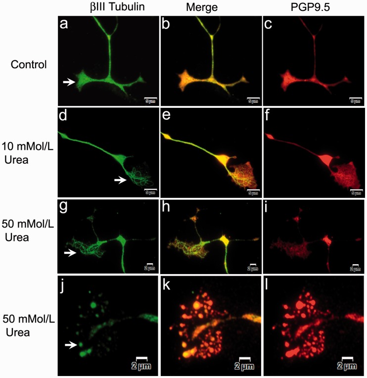
Figure 3.
βIII tubulin and PGP9.5 expression in growth cones. Widefield immunofluorescence showing growth cones of vehicle-treated neurons with uniformly distributed βIII tubulin (arrow in a, green), appearing yellow in the merged image (b) and overlapping the PGP9.5 expression (c, red). After 48 h 10 mmol/L urea treatment, neurons show growth cones containing distinct thickened βIII tubulin-positive fibres (d, green), with the merged image in (e), and pale depleted PGP9.5 expression (f, red). Similarly, 50 mmol/L urea-treated neurons show thickened individual βIII tubulin-positive fibres in the growth cone (g, green), merged image (i, yellow) and pale depleted PGP9.5 (h, red). Vesiculated remnants of the degenerating growth cone contain βIII tubulin (j, green), merged image (k, yellow) and PGP9.5 (l, red). Bar (a) to (f) = 5 µm, (g) to (l) = 2 µm.

