Figure 1.
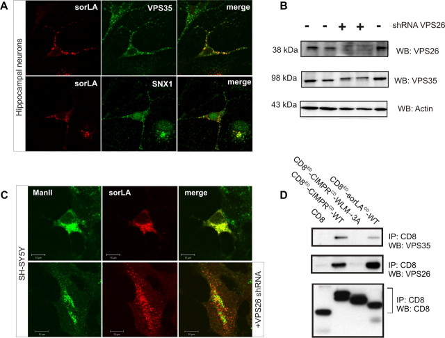
Retromer physically interacts with sorLA. A, Immunostaining of primary hippocampal neurons for sorLA (red) and VPS35 (in green; top) or SNX1 (in green; bottom). B, WB analysis of lysates from SH-SY5Y cells after transfection with (+) or without (−) an shRNA vector against human VPS26. Actin is used as loading control. C, SH-SY5Y cells were transfected with sorLA–WT (top) or sorLA–WT together with the VPS26 shRNA knockdown vector (bottom) sorLA (red) and the Golgi marker ManII (green). D, Co-IP analysis of CD8 chimeric proteins as previously (reported by Skinner and Seaman, 2009). Precipitated proteins were analyzed by WB.

