
Figure 4.
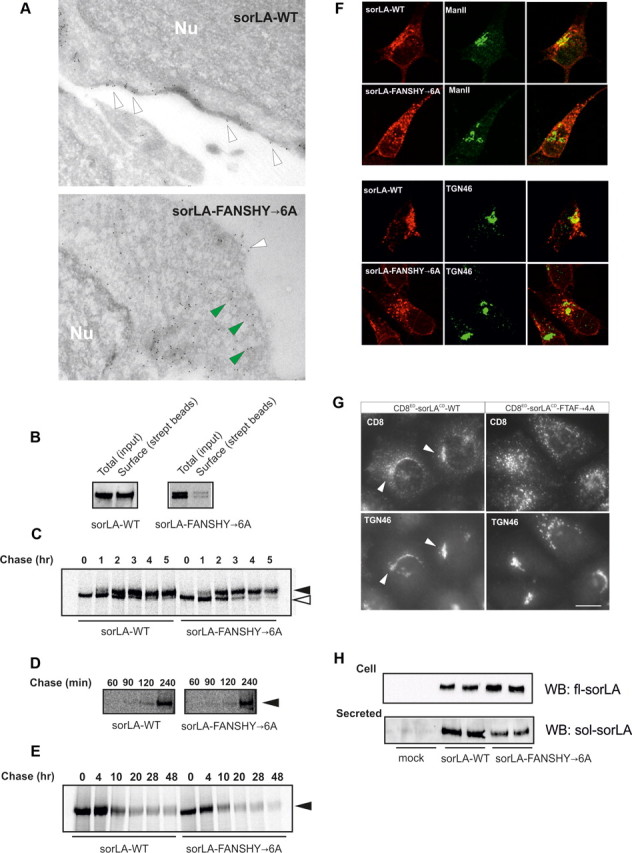
Characterization of sorLA transport in SH-SY5Y cell lines. A, Immunoelectron microscopy of sorLA–WT and sorLA–FANSHY→6A. Nu, Nucleus. Arrowheads in white indicate surface labeling and mutant receptors in the vicinity of the plasma membrane by green arrowheads. B, The level of biotinylated receptors at the cell surface (streptavidin beads) relative to the total expression level (input) in SH-SY5Y cells shown by representative WB. C, Pulse-chase experiment after the maturation of sorLA–WT and sorLA–FANSHY→6A in SH-SY5Y cells. Their glycosylation patterns are indicated by white (immature) and black (mature) arrowheads. D, Pulse-chase experiment of secreted sorLA ectodomains. E, Stability assay using pulse-chase technique. Proteins in C–E were visualized by radiography. F, SH-SY5Y cells stably expressing sorLA–WT or sorLA–FANSHY→6A were stained for the receptor (in red) together with markers of Golgi (ManII) or TGN (TGN46) (in green). G, Anti-CD8 antibody uptake in HeLa cells transfected with CD8ED–sorLACD–WT or CD8ED–sorLACD–FTAF→4A. Endosome-to-Golgi retrieval was evaluated by colocalization with TGN46. H, Secretion of sorLA extracellular domains at steady state determined by WB analysis of 45 h conditioned medium (Secreted) from SH-SY5Y cells that express sorLA–WT or sorLA–FANSHY→6A (Cell).