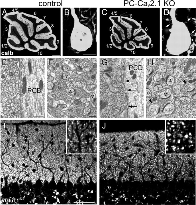
Figure 1.

Enlargement of PF terminals and proximal expansion of PF territory in PC-Cav2.1 KO mice. A–D, Immunofluorescence for calbindin (calb) in control (A, B) and PC-Cav2.1 KO mice (C, D). Arrows in D indicate spine-like protrusions from PC soma. The lobule number is indicated by numerals 1–10. E–H, Electron micrographs showing shaft dendrites of PCs (PCD; E, G) and neuropil regions (F, H) in control (E, F) and PC-Cav2.1 KO (G, H) mice. Arrows in G indicate ectopic spines from shaft dendrites. Asterisks in F and H indicate PC spines in contact with putative PF terminals. Arrowheads indicate PC spines thoroughly surrounded by enlarged PF terminals. I, J, Immunofluorescence for VGluT1 in control (I) and PC-Cav2.1 KO (J) mice. Scale bars: A, 500 μm; B, inset in I, 10 μm; E, F, 500 nm; I, 50 μm.
