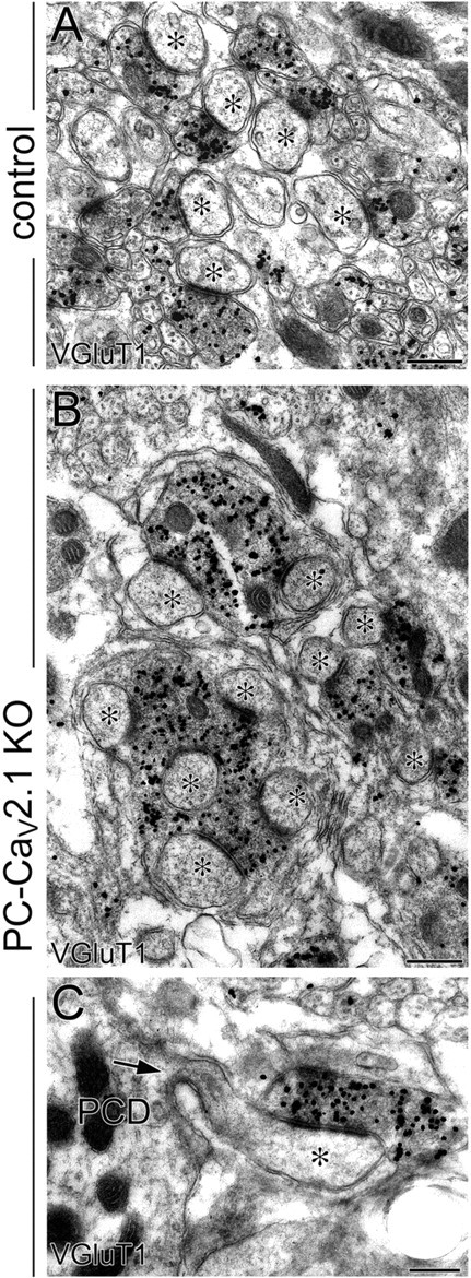
Figure 2.

Multiple spine contact with enlarged PF terminals and proximal innervation of PFs in PC-Cav2.1 KO mice. Preembedding immunogold microscopy for VGluT1 in control (A) and PC-Cav2.1 KO (B, C) mice. Asterisks indicate PC spines contacting to VGluT1(+) PF terminals. Arrow indicates a spine from PC dendrite (PCD). Scale bars, 500 nm.
