Figure 4.

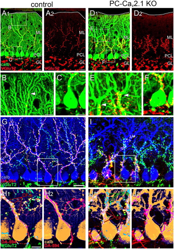
Regressed CF territory and multiple CF innervation in PC-Cav2.1 KO mice. A–F, Double immunofluorescence for calbindin (green) and VGluT2 (red) in control (A–C) and PC-Cav2.1 KO (D–F) mice. Boxed regions in A and D are enlarged in B, C, E, and F. Dotted lines indicate the pia mater. Arrowheads in B and E indicate the distal tips of VGluT2(+) CF terminals along PC dendrites. G–J, Triple fluorescent labeling for calbindin (blue in G, I; brown in H, J), VGluT2 (green), and anterograde tracer DA-594 injected into the inferior olive (red) in control (G, H) and PC-Cav2.1 KO (I, J) mice. Boxed regions in G and I are enlarged in H and J, respectively. calb, calbindin; CF-a and CF-b, tracer(+)/VGluT2(+) or tracer(−)/VGluT2(+) CFs, respectively; GL, granule cell layer; ML, Molecular layer; PCL, Purkinje cell layer. Scale bars: A, G, 20 μm; B, C, H, 10 μm.
