
Figure 7.
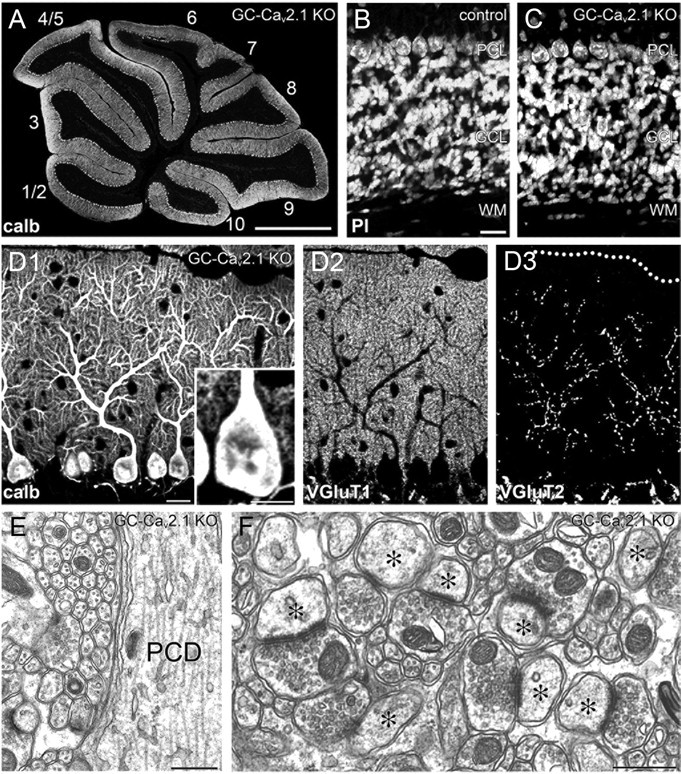
Normal synaptic wiring and structure in GC-Cav2.1 KO mice. A, Immunofluorescence for calbindin in the cerebellum of GC-Cav2.1 KO mice. The lobule number is indicated by numerals 1–10. B, C, Normal histology of the granular layer, as shown by propidium iodide (PI) staining in control (B) and GC-Cav2.1 KO (C) mice. D, Triple immunofluorescence for calbindin (D1), VGluT1 (D2), and VGluT2 (D3) in GC-Cav2.1 KO mice. E, F, Electron micrographs showing smooth surface of PC shaft dendrites (E) and normal morphology of PF–PC synapses (F). GCL, Granular cell layer; ML, molecular layer; PCD, Purkinje cell dendrite; PCL, Purkinje cell layer; WM, white matter. Scale bars: A, 1 mm; B, D1, 20 μm; D1, inset, 10 μm; E, F, 500 nm.