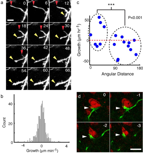
Figure 6.
Processes of multipolar cells are highly dynamic, grow in multiple directions, and make contact with mature neurons. a, Imaging time series of a retracting process (red arrowhead at tip) and a growing process (yellow arrowhead at tip). Time in minutes is indicated in the top right of each panel. Scale bar, 10 μm. b, Histogram of the length change rate for all the processes of four multipolar cells. Mean growth rate for all processes was not significantly different from 0 (t test, p = 0.625), suggesting that multipolar cells did not increase in size as they migrated but merely changed shape. c, On average, processes (blue circles) within 60° of the direction of soma movement grew during the hour before movement of the cell body, whereas processes oriented away from the direction of movement >60° retracted. Red dotted line corresponds to 0 μm growth per hour. d, Single optical confocal sections of a GFP+ process tip (green) making contact (arrowhead) with a NeuN+ soma (red) in HVC. Each section is separated by a 1 μm step; relative depth in micrometers is indicated on the top right of each panel. Scale bar, 10 μm.

