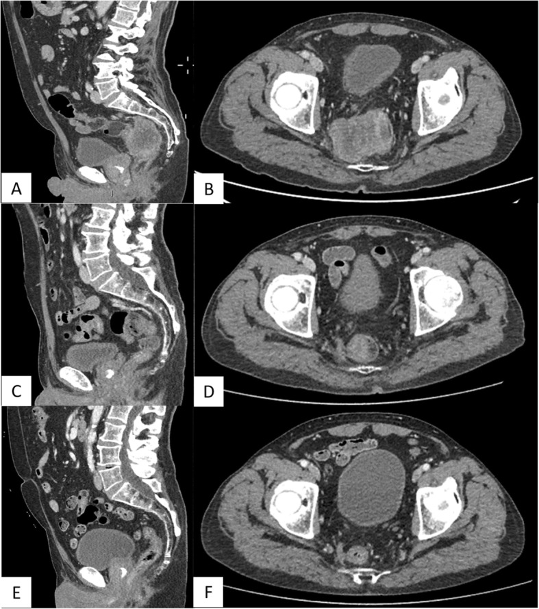
Fig. 1.

a, b CT scan showing a mass, centrally colliquated, originating from the right lateral wall of the rectum, infiltrating the right mesorectal fascia, the anterior presacral fascia, the homolateral piriformis muscle, and the right lobe of the prostate gland. The mass caused significant reduction in lumen calibre (a, sagittal plain. b, axial plain). c, d Re-evaluation of disease after the first three cycles of treatment. CT scan showed a marked reduction of the rectal mass of about 70–80%, with reduction also of lymph nodes and the prostatic involvement (c, sagittal plain. d, axial plain). e, f: CT evaluation after other three cycles of the same medical treatment. It showed further reduction of the rectal mass of about 50%. Lymph nodes and prostatic involvement disappeared (e, sagittal plain. f, axial plain)
