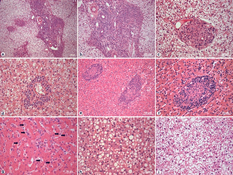
Fig. 1.
Histological changes in the liver of dogs with Leishmania infection. a, b Portal tract chronic inflammation. c Portal tract granuloma. d Perivascular inflammation. e, f Intralobular granuloma. g Hyperplasia and hypertrophy of Kupffer cells. h Steatosis. i Ballooning. Scale-bars: a, b, e, 100 µm; c, d, f–i, 50 µm

