Fig. 3.
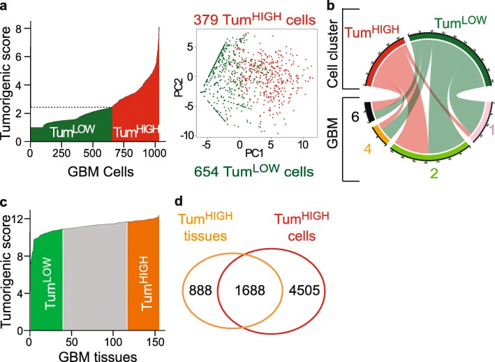
Signature-driven data reduction approach identifies cells according to their potential tumorigenic state. a Splitting cells into groups with high (TumHIGH) or low (TumLOW) tumorigenic potential. Left panel: tumorigenic score distribution across the cells. Dotted line: mean of the tumorigenic score. Right panel: PCA plot based on the expression of the 5 elements of the tumorigenic signature. b Contribution of each tumor to the two tumorigenic groups identified (chord plot). Note that each tumor contributes to each cell group. c Tumorigenic score distribution across GBM tissues (155 GBM tissues, TCGA RNA-seq dataset). TumHIGH and TumLOW GBM tissue groups selected at the extreme quartiles of the distribution. d High overlap between genes overexpressed in TumHIGH GBM tissues and cells. 65.5% (1688) of genes overexpressed in TumHIGH GBM tissues are also overexpressed in TumHIGH GBM cells

