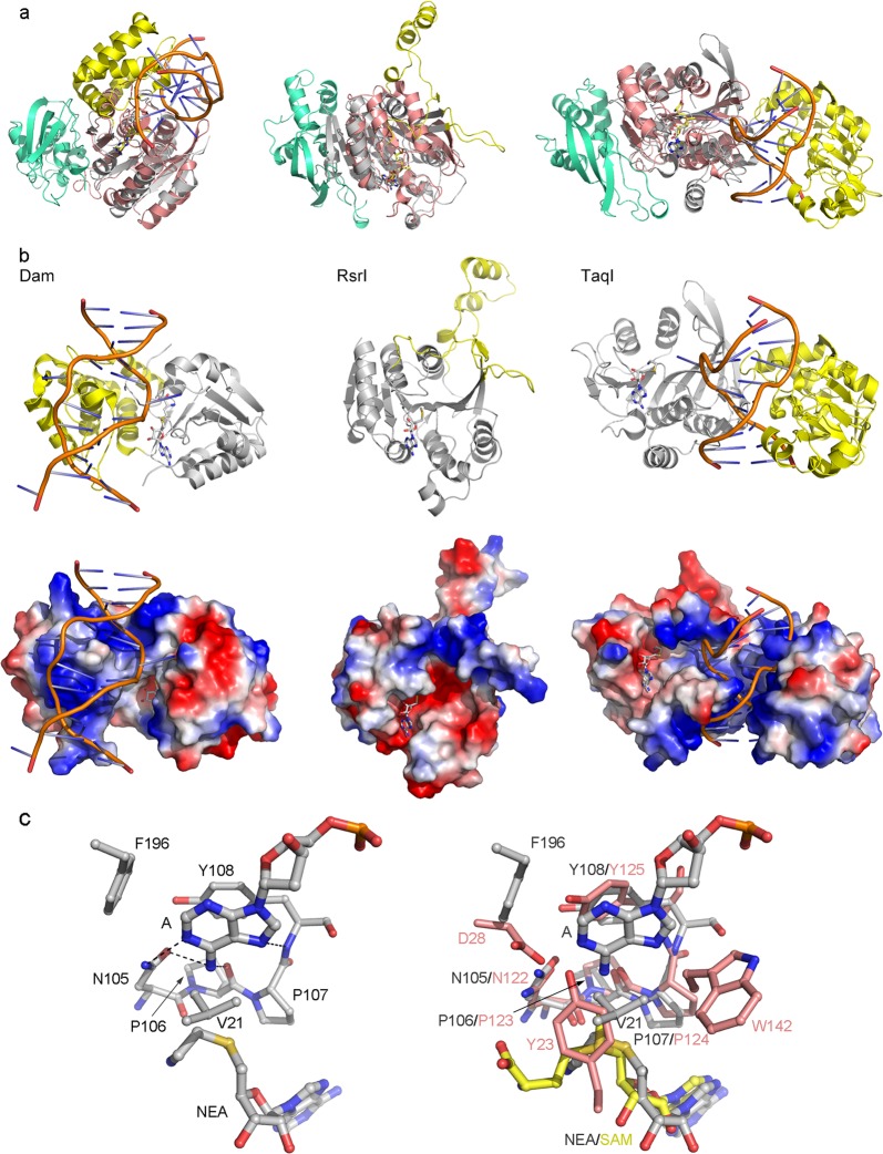
Fig. 4. Structural comparisons of human N6amt1–Trm112 complex with representative bacterial DNA 6mA MTases.
a Structural comparisons of N6amt1-Trm112 with Dam (left panel, PDB code 2G1P), RsrI (middle panel, PDB code 1EG2), and TaqI (right panel, PDB code 1G38). b Overall structures and Electrostatic potential surfaces of Dam, RsrI, and TaqI. The MTase and TRD domains of the bacterial DNA 6mA MTases are shown with ribbon models and colored in gray and yellow, respectively. The bound dsDNA is shown with orange ribbon. c Structure of the active site of TaqI (left panel) and comparison of the active site of N6amt1 with that of TaqI (right panel)

