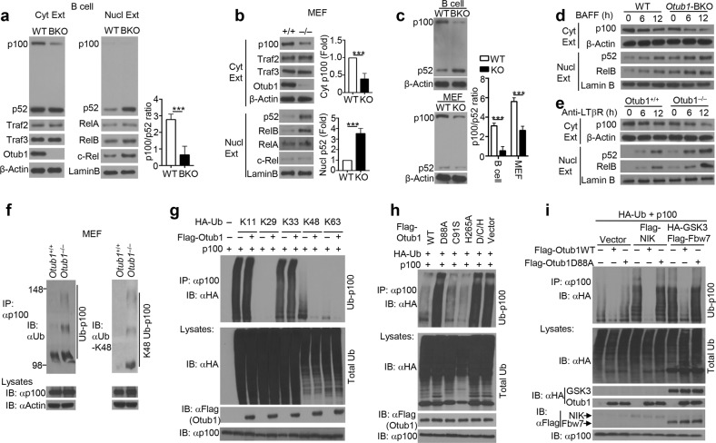
Fig. 2.
Otub1 regulates basal and signal-induced p100 processing and noncanonical NF-κB activation. a, b Immunoblot analysis of the indicated proteins using cytoplasmic (Cyt Ext) and nuclear (Nucl Ext) extracts of freshly isolated splenic B cells from wild-type (WT) and Otub1-BKO (BKO) mice (a) or Otub1+/+ (+/+) and Otub1–/– (–/–) primary MEFs (b). The p100 and p52 bands were quantified by densitometry based on three independent experiments and presented as the ratio of cytoplasmic p100 to nuclear p52 (a) or fold of cytoplasmic p100 and nuclear p52 with wild-type value being set to 1 (b). c Immunoblot analysis of p100 and p52 in whole-cell lysates of wild-type B cells or MEFs. The p100 and p52 bands were quantified by densitometry based on three independent experiments and presented as the ratio of p100 to p52 (right panel). d, e Immunoblot analysis of the indicated proteins using cytoplasmic or nuclear extracts of BAFF-stimulated splenic B cells (d) or anti-LTβR-stimulated primary MEFs (e). f Whole-cell lysates were prepared from MG132-treated (2 h) Otub1+/+ and Otub1–/– MEFs and subjected to p100 IP followed by detecting ubiquitinated p100 using antibodies detecting total ubiquitin chains (left) or K48-linked ubiquitin chains (right). The level of p100 and Actin in lysates was monitored by immunoblot. g p100 ubiquitination (upper panel) and direct immunoblot (lower panels) assays using whole-cell lysates of MG132-treated (for 2 h) HEK293 cells transfected with the indicated HA-tagged ubiquitin mutants (expressing only the indicated lysine with all other lysines mutated to arginines), either in the presence (+) or absence (–) of Flag-Otub1 and p100. h, i p100 ubiquitination (upper panel) and direct immunoblot (lower panels) assays using whole-cell lysates of MG132-treated (for 2 h) HEK293 cells transfected as indicated. D/C/H stands for Otub1 D88A/C91S/H265A mutant

