Fig. 5.
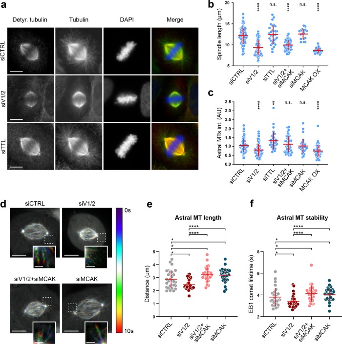
Vasohibins-mediated MT detyrosination regulates mitotic spindle length and positioning. a Representative images of mitotic spindles in control, VASH1/2- or TTL-depleted U2OS cells, immunostained with antibodies against detyrosinated and total α-tubulin. DNA was counterstained with DAPI. Detyrosinated tubulin in red, total α-tubulin in green, and DNA in blue in merged image. Scale bar: 10 µm. b Quantification of the spindle length from immunostained U2OS cells that were subjected to indicated treatments. c Astral MT intensities quantified as described in methods section. d Temporal color-coded projections of mitotic spindles of U2OS EB1-GFP cells treated with indicated siRNAs. Pronounced effect of the siRNA treatments on growing astral MTs shown in insets. Scale bar: 10 µm (Inset scale bar: 2 µm) e, f Astral MT length and stability quantified by manual tracking of EB1-GFP comet signal. N (number of cells, number of independent experiments): -spindle length: siCTRL (94, 6); siVASH1/2 (82, 5); siTTL (31, 2); siVASH1/2 + siMCAK (50, 3); siMCAK (32, 2); MCAK OX (49, 3); -astral MT intensity: siCTRL (106, 4); siVASH1/2 (73, 4); siTTL (33, 3); siVASH1/2 + siMCAK (51, 3); siMCAK (32, 2); MCAK OX (46, 3), -EB1-GFP comet tracking: siCTRL (29, 3); siVASH1/2 (27, 3); siVASH1/2 + siMCAK (29, 3); siMCAK (28, 3). P-values were calculated using Mann–Whitney U test. n.s. not significant, *p < 0.05, **p < 0.01, ****p < 0.0001

