Fig. 1.
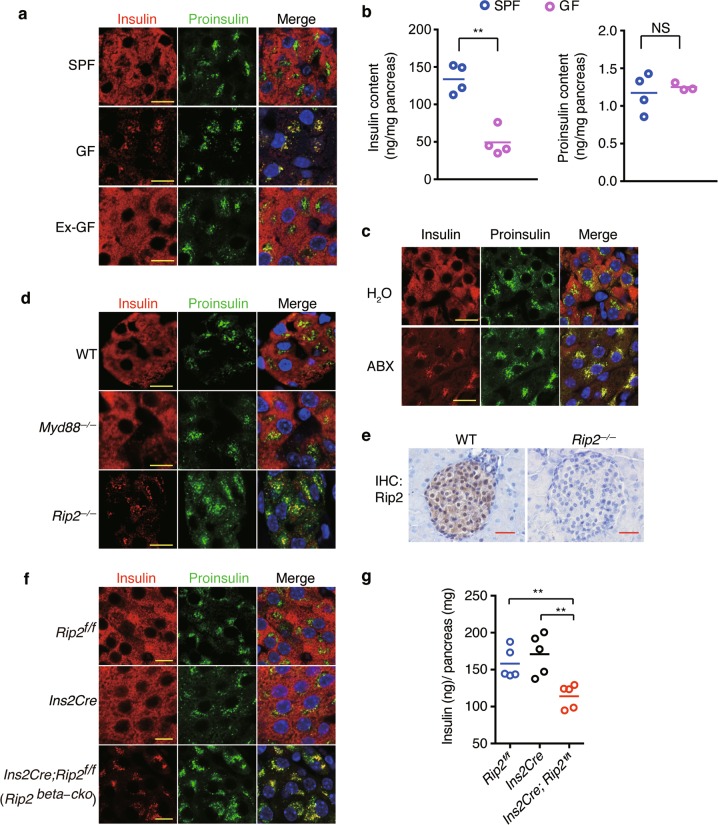
Beta cells sense microbes to direct insulin distribution in a cell-autonomous manner. a Immunostaining and confocal imaging of insulin (red) and proinsulin (green) in paraffin sections of pancreata from SPF, GF, and ex-GF mice. b The amount of insulin and proinsulin in pancreatic tissues from SPF and GF mice. c Immunostaining and confocal imaging of insulin (red) and proinsulin (green) in paraffin sections of pancreata from H2O (vehicle)- or antibiotic cocktail (ABX)-treated mice. d Immunostaining and confocal imaging of insulin and proinsulin in paraffin sections of pancreata from wild-type (WT), myd88−/− and Rip2−/− mice. e Immunohistochemical staining (IHC) of Rip2 in paraffin sections of pancreata from WT and Rip2−/− mice. f Immunostaining and confocal imaging of insulin and proinsulin in paraffin sections of pancreata from Rip2f/f, Ins2Cre, and Rip2beta-cko mice. g The amount of insulin in pancreatic tissues from Rip2f/f, Ins2Cre, and Rip2beta-cko mice. Nuclei were counter-stained in blue (a, c, d–f). Scale bars, 10 μm in a, c, d, f, 50 μm in e. Each symbol represents an individual animal, and horizontal bars indicate median values (b, g). P values were calculated with a two-tailed Student’s t-test (b) and a one-way ANOVA (g). **P < 0.01, NS, P > 0.05. Data (a–f) are representative of three independent experiments

