Fig. 4.
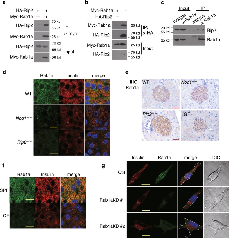
Nod1 and Rip2 recruit Rab1a onto DCVs to direct insulin vesicle transport. a, b Co-immunoprecipitation (IP) of Rip2 and Rab1a overexpressed in HEK293T cells. Tagged proteins were expressed in HEK293T cells, IP with anti-myc (a) or anti-HA (b) beads, and immunoblotted. c Co-IP of endogenous Rip2 and Rab1a in INS-1 cells. Rab1a was precipitated with an anti-Rab1a antibody. Rip2 and Rab1a were immunoblotted in input and IP fractions. d Immunostaining and confocal imaging of Rab1a and insulin in paraffin sections of pancreata from WT, Nod1−/− and Rip2−/− mice. e IHC staining of Nod1 or Rip2 in paraffin sections of pancreata from WT, Nod1−/−, Rip2−/−, and GF mice. f Immunostaining and confocal imaging of Rab1a and insulin in paraffin sections of pancreata from SPF and GF mice. g Immunostaining and confocal imaging of insulin and Rab1a in INS-1 cells with stable shRNA knockdown of Rab1a. Ctrl, scramble shRNA; Rab1aKD #1 or Rab1aKD #2, shRNAs against Rab1a. Nuclei were counter-stained in blue (d–g). Scale bars, 50 μm (e), 10 μm (d, f, g). Data are representative of three independent experiments

