Figure 2.
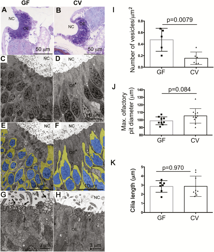
Ultrastructural and cellular changes in the zebrafish olfactory organ in the absence of microbiota. (A) Light micrograph of a semithin section of a GF zebrafish olfactory organ stained in toluidine blue. (B) Light micrograph of a semithin section of a CV zebrafish olfactory organ stained in toluidine blue. (C, E, and G) Transmission electron micrograph (TEM) of a GF zebrafish olfactory epithelium. (D, F, and H) TEM of a CV zebrafish olfactory epithelium. Images in (E) and (F) were pseudocolored as explained in the materials in methods to highlight the nucleus area (blue) and the cytoplasm (yellow) of OSNs. Arrows in (G) indicate transport vesicles in the apical pole of sustentacular cells. NC: nasal cavity; OE: olfactory epithelium. (I) Number of apical vesicles quantified in TEM images from CV and GF zebrafish larvae (N = 3 larvae/group). (J) Mean maximum olfactory pit diameter measured in CV and GF zebrafish larvae (N = 10). (K) Quantification of the maximum ciliar length in CV and GF zebrafish (N = 8).

