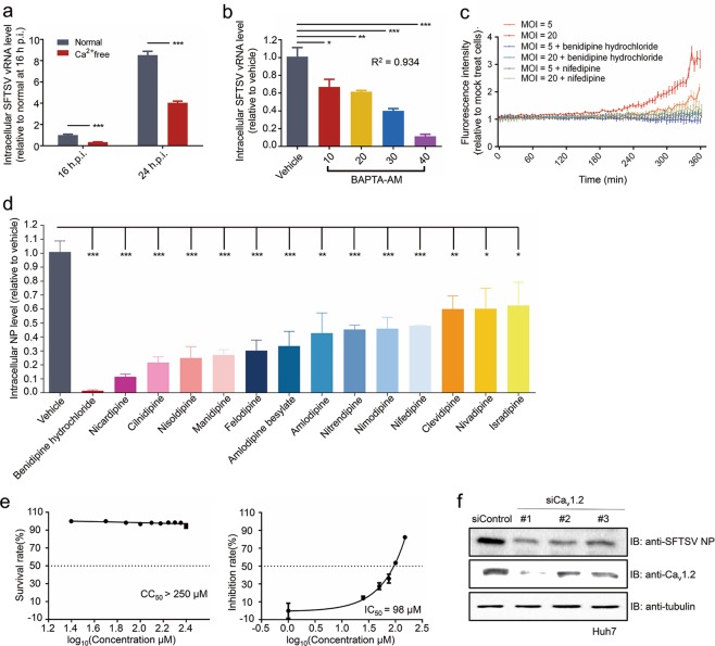
Fig. 3.
In vitro anti-SFTSV activity of CCBs. a Vero cells were cultured in Ca2+-free or normal medium and then infected with SFTSV (MOI = 1). At 16 or 24 hpi, relative intracellular SFTSV vRNA level was measured by qRT-PCR. b Relative intracellular vRNA level of SFTSV in Vero cells upon treatment with BAPTA-AM at 24 hpi. c Intracellular Ca2+ concentration in SFTSV-infected cells. Vero cells were infected with SFTSV at the indicated MOI in the presence of benidipine hydrochloride or nifedipine or vehicle (DMSO). Relative intracellular Ca2+ level was determined by fluorescence of Fluo-4NW. d Relative intracellular SFTSV NP level in the infected Vero cells. Vero cells were treated with the indicated 14 CCBs or vehicle (DMSO), infected with SFTSV at MOI of 1 and the relative NP level was determined by immunofluorescence with NP antibody at 36 hpi. e Survival rate analysis (left) and dose-dependent inhibition effects (right) of nifedipine treatment in Vero cells. f Relative intracellular SFTSV NP level in SFTSV-infected Huh7 cells. Cells were transfected with siRNA against L-type calcium channel Cav1.2 for 48 h and then infected with SFTSV for 48 h, followed by western blot analysis of intracellular NP level. Comparison of mean values (a, b, and d) between two groups was analyzed by Student’s t-test. *P < 0.05; **P < 0.01; ***P < 0.001

