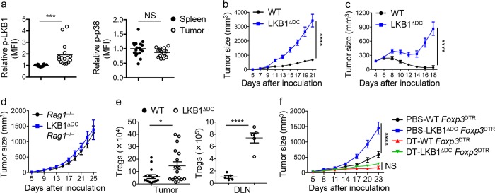
Fig. 1.
LKB1 is activated in DCs in tumor, and its deletion in DCs impairs anti-tumor immunity. a C57BL/6 mice were inoculated with MC38 tumor cells for 14 days and DCs from tumor tissues and spleen were analyzed for p-LKB1 and p-p38 expression. b, c Tumor growth curve in wild-type (WT) and LKB1ΔDC mice following inoculation of MC38 tumor cells (b; WT, n = 12; LKB1ΔDC mice, n = 9) or MC38-EGFR cells (c; WT, n = 8; LKB1ΔDC mice, n = 9). d Sublethally irradiated Rag1−/− mice were reconstituted with control Rag1−/−- or LKB1ΔDCRag1−/−-derived bone marrow cells for chimera generation, and subsequently inoculated with MC38 tumor cells for tumor growth curve. e Statistics of Treg numbers in tumor tissues and draining lymph nodes (DLN) of WT and LKB1ΔDC mice after inoculation of MC38 tumor cells for 13–14 days. f Sublethally irradiated Rag1−/− mice were reconstituted with control Foxp3DTR- or LKB1ΔDCFoxp3DTR-derived bone marrow cells for chimera generation, and subsequently treated with or without diphtheria toxin (DT), and inoculated with MC38 tumor cells for tumor growth curve (n = 10 per group). Data in plots indicate the means ± s.e.m; each symbol represents an individual mouse. NS not significant; *P < 0.05, ***P < 0.001, ****P < 0.0001; two-tailed unpaired Student’s t-test (a, e) or two-way ANOVA (b, c, f). Data are from three (a, b, e) or two (c, d, f) independent experiments

