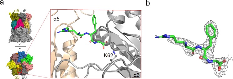
Figure 9.
Interaction of 6 with yeast 20S proteasome (PDB code 4X6Z). (a) General (left) and detailed (blow-up) localization of the inhibitor binding site between α5 (wheat color) and α6 (gray) subunits. It is visible in the crystal structure that the C-terminal fragment of 6 (green) binds to the ε-amine group of the conserved α6Lys62 through its carboxylate group. (b) Electron density defining the fragment of 6 included in the model (2Fo – Fc omit map contoured at 1σ level). The cartoon of general proteasome structure in (a), left, has also been used in our earlier work.30

