
FIG 2.
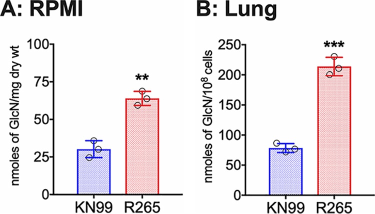
C. gattii R265 cells produce significantly larger amount of chitosan in the cell wall compared to C. neoformans under host conditions. (A) Chitosan levels of strains grown in RPMI 1640 containing 10% FBS and 5% CO2 at 37°C for 5 days. Strains were grown in YPD for 2 days. Yeast cells were harvested, washed with PBS, inoculated at 500 cells/μl in RPMI 1640 containing 10% FBS, and incubated for 5 days at 37°C in the presence of 5% CO2. At the end of incubation, chitosan was measured by the MBTH assay and expressed as nanomoles of glucosamine per milligram (dry weight) of cells. Data represent the averages from three biological experiments. (B) Chitosan levels of strains growing in the murine lung. Mice (CBA/J) (three mice per group) were intranasally inoculated with 107 CFU of each strain. On day 7 postinfection (PI), the lungs were excised and homogenized, and the lung tissue was removed by alkaline extraction, leaving the fungal cells to be harvested, counted, and subjected to the MBTH assay. Data are expressed as nanomoles of glucosamine per 108 cells. Significant differences between the groups were compared by two-tailed unpaired t test with Welch’s correction. Error bars represent standard errors of the means. ***, P < 0.0062; **, P < 0.001.