FIG 2.
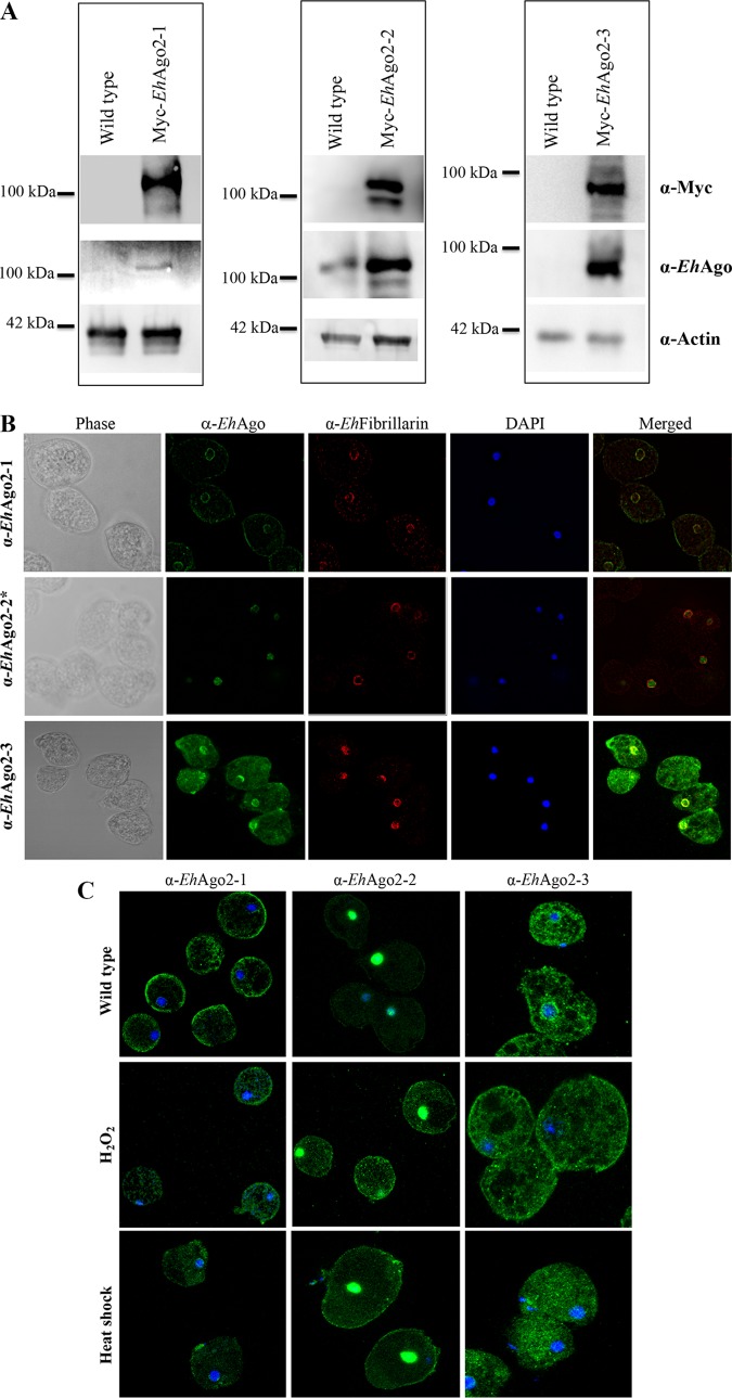
IFAs for the three EhAgos show distinct subcellular localization in the parasite, and stress conditions affect their expression/localization. (A) Custom-made antibodies to the three EhAgos detect specific bands by Western blotting. Western blot analyses were performed on total cell lysates from wild-type and Myc-tagged overexpressing cell lines. The expected band size of EhAgo2-2 was detected in both lysate sample lanes; however, EhAgo2-1 and EhAgo2-3 can be detected only in lysates from overexpressing cell lines and not in wild-type cell lysates, likely due to their low endogenous expression, based on three published data sets using Affymetrix microarray (24, 25) or RNA-seq (26). Anti-actin was used as a loading control. (B) Distinct subcellular localizations are shown for each EhAgo in the parasite by IFAs. E. histolytica trophozoites were fixed and immunostained using custom peptide antibodies for EhAgo2-1, EhAgo2-2, and EhAgo2-3. Localization for EhAgo2-2 has previously been published (23) and was used as a control. A ring structure at the perinuclear location was seen for both EhAgo2-1 and EhAgo2-3, contrasting with the nuclear staining for EhAgo2-2 IFA. Additional signal was seen for EhAgo2-1 (cell surface membrane) and for EhAgo2-3 (cytosol). Anti-EhFibrillarin is used as a marker of the nucleolus, which is located at the nuclear periphery in E. histolytica. DAPI, 4′,6-diamidino-2-phenylindole. (C) Localization of EhAgo proteins changes in response to stress conditions. IFAs of merged image of DAPI and each anti-EhAgo are shown for untreated parasites and parasites treated with two stress conditions (42°C for 1 h or 1 mM H2O2 for 1 h). Compared with the wild-type condition, the EhAgo2-1 localization of the perinuclear ring was almost lost under H2O2 treatment but was largely unchanged with heat shock. The EhAgo2-2 signal completely overlapped with the DAPI signal in untreated parasites but was greatly increased throughout the cytoplasm under both stress conditions, along with noticeable punctate dots, but its nuclear localization signal remained for both stress conditions. The EhAgo2-3 perinuclear localization is no longer observed under both stress conditions, and the major signal of staining is from cytoplasm. All stress condition assays were performed with multiple biological replicates and gave reproducible results; representative images are shown.

