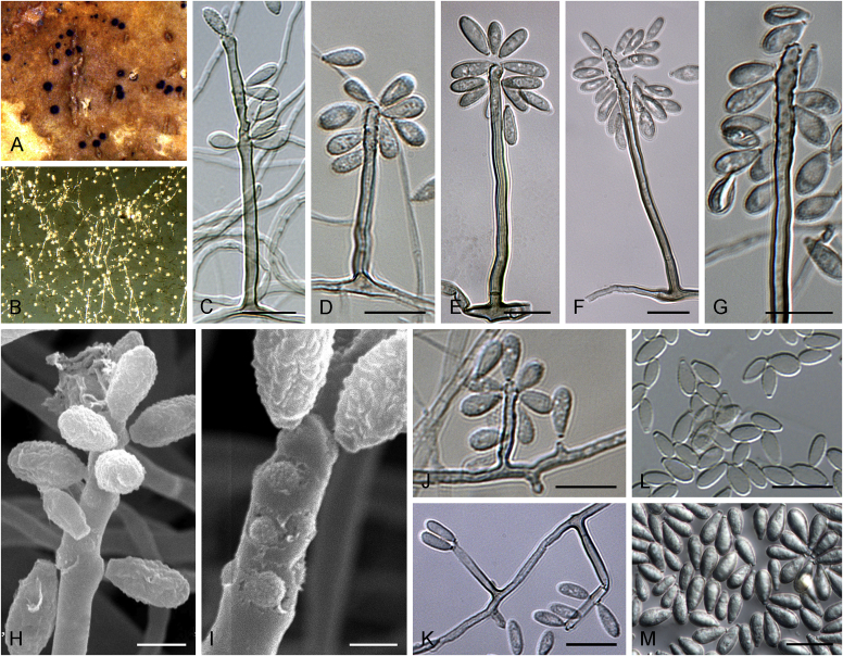
Fig. 59.
Ramichloridium spp. A.Ramichloridium luteum on apple. B. Sporulating colonies of Ramichloridium luteum (ex-type CBS 132088) on PDA. C–G. Macronematous conidiophores with sympodially proliferating conidiogenous cells, which give rise to a conidium-bearing rachis with crowded and prominent scars. C.Ramichloridium apiculatum (ex-type CBS 156.59). D.Ramichloridium cucurbitae (ex-type CBS 132087). E, F.Ramichloridium luteum (ex-type CBS 132088). G.Ramichloridium punctatum (ex-type CBS 132090). H, I. Scanning electron micrographs of Ramichloridium luteum (ex-type CBS 132088) showing sympodial proliferation with scars on conidiogenous cells. J, K. Conidiophores reduced to conidiogenous cells. J.Ramichloridium cucurbitae (ex-type CBS 132087). K.Ramichloridium luteum (ex-type CBS 132088). L, M. Conidia. L.Ramichloridium apiculatum (ex-type CBS 156.59). M.Ramichloridium punctatum (ex-type CBS 132090). Scale bars: H = 2 μm; I = 1 μm; all others = 10 μm. Pictures C, L taken from Li et al. (2012); all others from Arzanlou et al. (2007).

