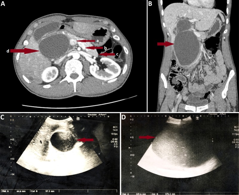
Figure 1. Pictures of the patient's preoperative CT scan and ultrasounds .
Figures 1A, 1B are axial and coronal images of preoperative CT scan of patient undertaken three weeks prior to admission, which depict the size and extent of the giant Todani type I choledochal cyst. In Figure 1A, a shows portal vein, b shows splenic vein, c shows body and tail of pancreas, and d shows the giant choledochal cyst almost completely replacing the head of the pancreas. In Figure 1B, the arrow shows the choledochal cyst in a coronal image. Figure 1C is an ultrasound image of the patient on the day of admission; the arrow shows the size of choledochal cyst. Figure 1D is an ultrasound image of the patient on the 7th-day post-admission after his condition suddenly deteriorated; the arrow shows the choledochal cyst at this point in time. In Figures 1C, 1D, the sudden increase in size on the 7th post-admission day can be appreciated.

