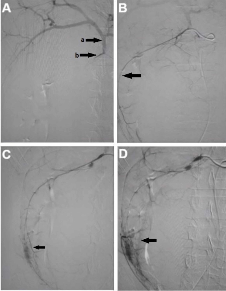
Figure 2. Serial images of attempted angioembolization of bleeding vessels of choledochal cyst during vascular intervention.
Figure 2A shows initial selective cannulation of gastroduodenal artery. a shows hepatic arterial system and b shows the origin of the gastroduodenal artery. Figure 2B shows the feeding vessel of the choledochal cyst arising from the gastroduodenal artery. Figures 2C, 2D show the blush representing bleeding into the choledochal cyst and confirming the diagnosis of arteriocholedochal fistula.

