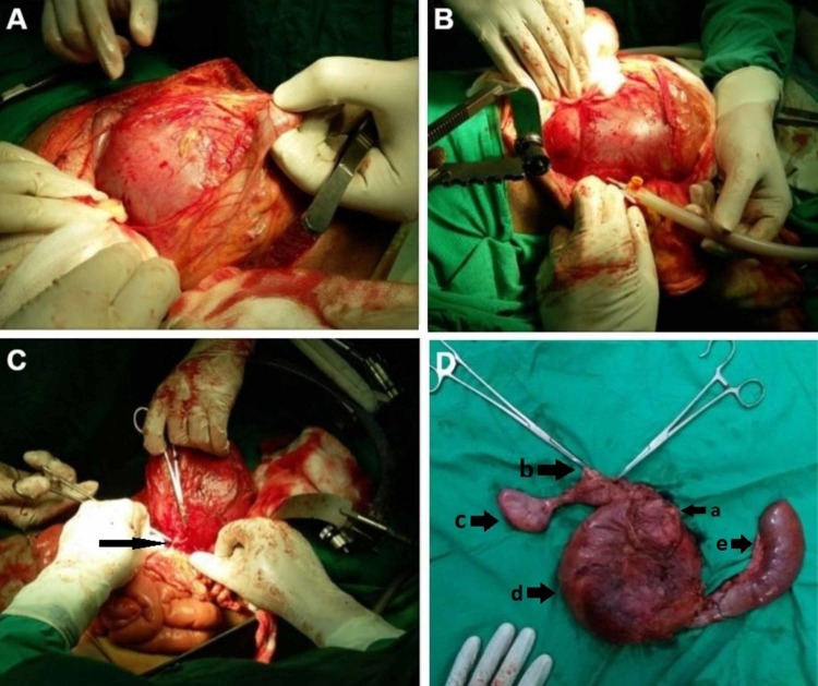
Figure 3. Peroperative pictures showing the giant choledochal cyst and final resected specimen.
Figure 3A shows the giant choledochal cyst upon opening the abdominal cavity. FIgure 3B shows aspiration of the giant choledochal cyst. Figure 3C shows the collapsed cyst with a feeding vessel being ligated that was arising from posterior to the pancreas, probably from a branch of the superior mesenteric artery. Figure 3D shows the final resected Whipple's pancreatoduodenectomy specimen. In Figure 3D, a shows the atrophied head of pancreas, b shows upper resection margin of the choledochal cyst, c shows the gallbladder and cystic duct, d shows collapsed choledochal cyst and e shows fourth part of the duodenum and jejunum

